
Muskan Taneja
5 min read
Introduction
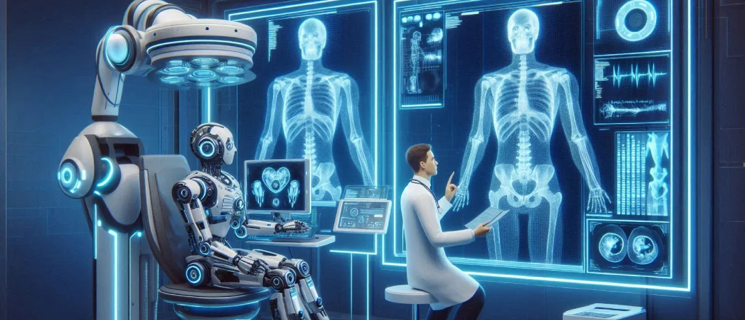
Artificial Intelligence is transforming many fields, and radiology is no exception. Radiology involves the use of medical imaging to diagnose and treat diseases within the body, and AI is helping to enhance the accuracy and efficiency of these processes. From improving diagnostic accuracy to streamlining workflow, AI is transforming how radiologists work. In this blog, we'll explore the benefits, use cases, real-life examples of AI in radiology and much more.
So, without further ado. Let’s get started.
Benefits of AI in Radiology
-
Enhanced Diagnostic Accuracy
AI algorithms, especially those based on deep learning, can analyze medical images with remarkable precision. These algorithms can detect subtle patterns that might be missed by the human eye, leading to earlier and more accurate diagnoses.
-
Increased Efficiency
AI can automate repetitive tasks such as image segmentation and analysis, allowing radiologists to focus on more complex cases. This not only speeds up the diagnostic process but also reduces the workload on healthcare professionals.
-
Improved Workflow
AI-powered systems can prioritize cases based on urgency, ensuring that critical cases are reviewed first. This helps in managing patient flow and reducing waiting times for urgent diagnoses.
-
Cost Reduction
By increasing efficiency and accuracy, AI can help reduce healthcare costs. Early and accurate diagnosis can lead to better patient outcomes and reduce the need for expensive treatments.
-
Enhanced Learning and Training
AI can be used to train radiologists by providing them with a vast number of annotated images to practice on. This helps in improving their diagnostic skills and keeping them updated with the latest developments in the field.
Use Cases of AI in Radiology
-
Enhancing Cardiac Imaging
AI algorithms enhance cardiac imaging by analyzing cardiac MRI and CT scans to assess heart function, detect structural abnormalities, and identify areas of ischemia or infarction. These tools improve diagnostic accuracy in cardiovascular diseases, reduce the time required for analysis, and support early intervention strategies. By providing detailed insights into cardiac health, AI helps in better management and treatment of heart conditions.
-
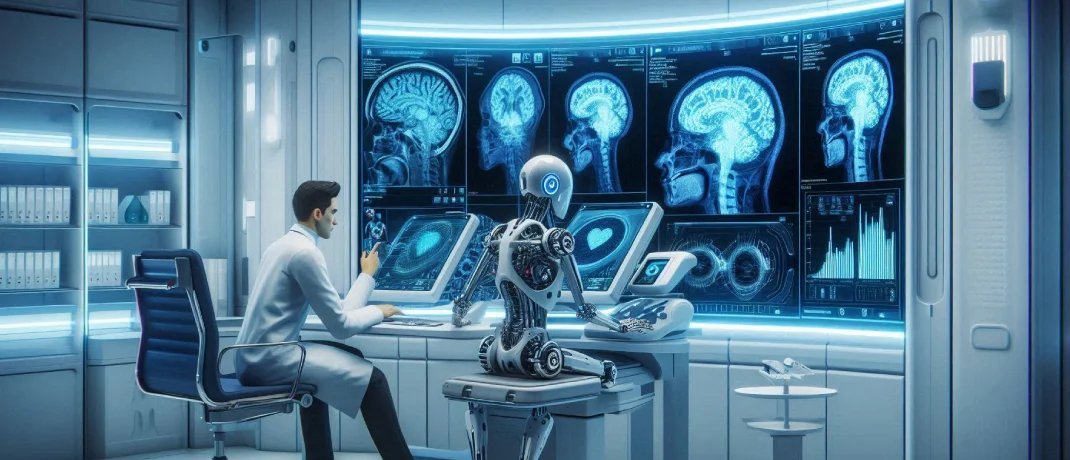
Classifying Brain Tumours
AI applications in radiology include classifying brain tumours by analyzing MRI scans. These algorithms can distinguish between different types of tumours, such as gliomas and meningiomas, with high accuracy. This capability enhances diagnostic precision, aids in personalized treatment planning, and monitors tumour progression or response to therapy, ultimately improving patient outcomes.
-
Spotting Vertebral Fractures
AI systems are adept at detecting vertebral fractures on spine X-rays and CT scans, even when fractures are subtle. This early detection is crucial for patients with osteoporosis or other conditions predisposing them to fractures. By identifying fractures that might be missed by human eyes, AI helps prevent complications and ensures timely intervention.
-
Detecting Alzheimer’s Disease
AI tools analyze brain imaging, including MRI and PET scans, to detect early signs of Alzheimer's disease. These tools can identify brain atrophy and other biomarkers indicative of the disease's onset. Early detection allows for timely therapeutic interventions and better management of the disease's progression, potentially improving the quality of life for patients.
-
Diagnosing ALS
In diagnosing Amyotrophic Lateral Sclerosis (ALS), AI can analyze neuroimaging data to detect patterns associated with the disease. Early and accurate diagnosis is challenging but crucial, and AI aids by identifying subtle changes in motor neurons and brain regions. This helps in starting treatments earlier, potentially slowing the disease's progression and aiding in patient care.
-
Assisting with Radiology Reporting & Data-Related Tasks
AI assists radiologists by automating reporting and handling data-related tasks. Natural language processing algorithms can generate accurate radiology reports, extract relevant patient data, and integrate findings from various imaging studies. This automation reduces administrative burdens, minimises errors, and allows radiologists to focus more on patient care and complex diagnostic tasks.
-
Detecting Breast Cancer
AI in mammography and breast MRI aids in the early detection of breast cancer by identifying suspicious lesions and microcalcifications with high precision. These tools improve screening accuracy, reduce false positives, and help in differentiating between benign and malignant findings. Early and accurate detection of breast cancer significantly enhances the chances of successful treatment and patient survival.
-
Dose Optimization
AI plays a vital role in dose optimization by adjusting radiation doses in imaging procedures to the minimum required levels without compromising image quality. This helps in reducing the risk of radiation exposure to patients, particularly in repeated imaging studies. AI-driven dose management ensures patient safety while maintaining diagnostic efficacy.
-
Detecting Pneumonia
AI algorithms can analyze chest X-rays and CT scans to detect pneumonia, including cases caused by COVID-19. These tools can quickly identify lung infections and differentiate between bacterial and viral causes. Early and accurate detection of pneumonia facilitates timely treatment, which is crucial for patient recovery, especially in severe cases.
-
Detecting LVO (Large Vessel Occlusion)
AI is used to detect large vessel occlusions in stroke patients by analyzing CT angiography and MRI scans. Rapid identification of LVO is critical for administering timely treatments such as thrombectomy. AI enhances the speed and accuracy of LVO detection, improving outcomes for stroke patients by enabling quicker intervention and reducing the risk of long-term disability.
Also Read: How to Build a Telemedicine App like Teladoc?
Real-life Examples of AI in Radiology
-
Google Health's AI for Breast Cancer Detection
Google Health has developed an AI model that can detect breast cancer with greater accuracy than human radiologists. In a study published in Nature, the AI reduced false positives by 5.7% and false negatives by 9.4% in the UK, demonstrating its potential to improve breast cancer screening.
-
Aidoc's AI Solutions for Radiology
Aidoc offers AI solutions that assist radiologists in detecting critical conditions, such as intracranial hemorrhages and pulmonary embolisms. Their AI algorithms analyze medical images in real-time and alert radiologists to urgent cases, enhancing the speed and accuracy of diagnosis.
-
Zebra Medical Vision's AI Radiology Assistant
Zebra Medical Vision has developed an AI radiology assistant that can detect a range of conditions, including liver disease, osteoporosis, and cardiovascular issues. Their algorithms have been trained on millions of medical images, enabling them to provide accurate and reliable diagnoses.
-
IBM Watson Health's Imaging AI
IBM Watson Health uses AI to analyze medical images and provide insights into various conditions, such as cancer and cardiovascular diseases. Their AI-powered tools help radiologists in making more informed decisions and improving patient outcomes.
-
Arterys' AI for Cardiac Imaging
Arterys has developed AI solutions for cardiac imaging that can analyze MRI scans and provide detailed assessments of heart function. Their AI algorithms can perform tasks such as measuring ejection fraction and detecting cardiac anomalies, aiding in the diagnosis and treatment of heart diseases.
Challenges of AI in Radiology
-
Data Quality and Quantity
AI models in radiology require extensive, high-quality annotated data for effective training. However, obtaining such data is challenging due to privacy concerns and the variability in imaging practices across different institutions. Additionally, the availability of annotated datasets is limited, which can lead to biased or less effective AI models. Ensuring consistent data quality and sufficient quantity remains a significant hurdle in the development and deployment of AI solutions in radiology.
-
Integration into Clinical Workflows
Integrating AI tools into existing clinical workflows is complex, necessitating substantial changes in how radiologists perform their tasks. This integration can be met with resistance from healthcare professionals who may be wary of altering established practices. Moreover, disruptions caused by the introduction of new technologies can hinder the adoption and effective use of AI. Ensuring seamless integration while maintaining efficiency is a critical challenge.
-
Regulatory and Ethical Issues
AI applications in radiology must navigate stringent regulatory requirements and address ethical concerns, particularly related to patient data privacy and algorithmic bias. The regulatory landscape for AI in healthcare is still evolving, leading to uncertainties that can delay deployment. Ethical considerations, such as ensuring unbiased algorithms and protecting patient confidentiality, are paramount and add layers of complexity to the adoption process.
-
Interpretability and Trust
Many AI models, especially those based on deep learning, function as "black boxes," providing results without clear explanations of how decisions are made. This lack of interpretability can lead to mistrust among radiologists, who may be hesitant to rely on AI without understanding its decision-making process. Enhancing the transparency and explainability of AI models is crucial to gaining the trust and acceptance of healthcare professionals.
-
Continuous Learning and Adaptation
Medical imaging technologies and practices are continually evolving, necessitating that AI models also adapt to new data and protocols. This requirement for continuous learning and adaptation is technically challenging and resource-intensive. Maintaining the relevance and accuracy of AI systems in the face of ongoing advancements in radiology is a persistent challenge that requires robust mechanisms for real-time updates and model retraining.
Also Read: A Comprehensive Guide to Healthcare Software Development in 2024
Future Directions of AI in Radiology
-
Improved Data Sharing and Collaboration
The establishment of secure, interoperable data-sharing platforms will enable the aggregation of large, diverse datasets from multiple institutions. This collaboration will enhance the quality and generalizability of AI models by providing richer data for training and validation. Improved data sharing can help reduce biases, improve model performance, and foster innovation in AI development within radiology.
-
Seamless Integration with PACS and EHR Systems
Developing AI solutions that integrate seamlessly with Picture Archiving and Communication Systems and Electronic Health Record systems will streamline workflows and enhance clinical utility. Such integration will facilitate the smooth adoption of AI tools, allowing radiologists to incorporate AI-driven insights directly into their diagnostic processes, thereby improving efficiency and accuracy.
-
Regulatory Frameworks and Standards
The establishment of clear regulatory frameworks and industry standards for AI in radiology is essential to ensure the safety, efficacy, and ethical use of these technologies. Regulatory clarity and standardized guidelines will help accelerate the approval and implementation of AI tools in clinical practice. This will provide a structured pathway for developers and increase confidence among healthcare providers and patients.
-
Enhancing Interpretability and Explainability
Research and development efforts focused on making AI models more interpretable and explainable are crucial for building trust among radiologists and healthcare providers. Improved interpretability will allow radiologists to understand and validate the AI's decision-making process, fostering better collaboration between human expertise and AI capabilities. This will lead to greater acceptance and reliance on AI-assisted diagnostics.
-
Continuous Model Learning and Real-Time Updates
Implementing mechanisms for continuous learning and real-time updates will enable AI models to remain current with the latest medical imaging techniques and practices. These capabilities will ensure that AI systems can adapt to new data and evolving clinical protocols, maintaining their relevance and accuracy over time. Continuous model learning will be essential for keeping AI tools effective and reliable in dynamic healthcare environments.
Conclusion
In conclusion, AI is revolutionizing radiology by improving diagnostic accuracy, streamlining workflows, and enhancing patient outcomes. Its applications range from detecting diseases like breast cancer and Alzheimer's to automating routine tasks and optimizing imaging processes. Despite challenges such as data quality and integration into clinical workflows, the future of AI in radiology is promising, with advancements in data sharing, regulatory frameworks, and model transparency paving the way for further innovations. As AI continues to evolve, it will play an increasingly vital role in transforming radiology and healthcare overall.
